


(fibroelastometry) of the liver
In most patients, viral hepatitis C takes on a chronic course after a short-term acute onset. For many years, the hepatitis C virus (HCV) has been present in the liver tissue (parenchyma), and changes its structure. Analysis of fibroelastometry (syn. - elastometry) helps to detect these structural changes.
What is this analysis (why do it)
This type of hardware diagnostics for hepatitis C is carried out together with other studies. According to the results of elastometry, one can judge how much the parenchyma has suffered in hepatitis C. Fibroelastometry of the liver - what is it, and what is its price? The hepatic parenchyma consists of cells, hepatocytes.
These cells are the breeding ground for the virus. HCV enters the hepatocytes and multiplies within them. As a result, a significant part of these cells die. In place of the dead hepatocytes, fibrous connective tissue grows.
Hepatic fibrosis progresses. With an extreme degree of fibrosis, cirrhosis, the amount of parenchyma that has become is minimal. Clinically, this is manifested by liver failure. Fibroelastometry helps:
- detect fibrosis in time
- determine its degree
- diagnose cirrhosis
- determine the duration of AVT (antiviral therapy)
- make a prediction about the outcome of the disease
- monitor the effectiveness of treatment (through repeated studies).
The essence of this research is based on such physical quantities as elasticity and stiffness. Both of these values will change with hepatic fibrosis. A healthy liver is more elastic in comparison with a diseased liver. Under the action of an external force, it is deformed, and after the termination of the force action, it quickly takes on its original shape. With fibrosis, this process slows down.
Rigidity is the ability to withstand external force and retain its original shape under its influence. Stiffness is inversely proportional to elasticity. The stronger the fibrosis, the greater the stiffness. A healthy liver without fibrosis is as elastic as possible. And the liver with cirrhosis is as hard as possible.
Diagnostics is carried out using the FibroScan apparatus. This machine is equipped with a high-resolution convex probe. This broadband transducer emits ultrasonic waves with a frequency of 2.5 MHz and high penetration.
When passing through internal media, the characteristics of ultrasonic waves change. These changes are processed by the device and displayed graphically on the monitor, as well as in the form of numerical indicators. The Fibroscan device is so popular with doctors and patients that its name has become a household name. Fibroelastometry of the liver is often called fibroscan.
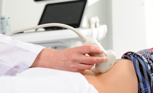
How is the procedure
Diagnostics is carried out in a specialized office equipped with Fibroscan equipment. The patient lies down on a flat hard surface of the couch, having previously undressed to the waist or exposing the abdomen. To ensure that the results are not distorted, a close contact of the sensor with the patient's body is required. To do this, the doctor treats the skin of the abdominal wall in the projection with a special gel, and then proceeds to the study. It is carried out in two forms.
The first type is compression or quasi-static elastometry. The sensor is located in the projection of the right lobe of the liver, in the VI-VIII intercostal space along the mid-axillary line. In this position, the doctor makes compression, squeezing, or a series of squeezing of soft tissues. The compression vector should be directed perpendicular to the surface of the organ.
The deformation should not exceed 1-2%. New modifications of Fibroscan devices are equipped with sensors with devices that generate mechanical compression pulses with preset values. Thanks to such devices, errors due to incorrect execution technique are minimized.
The second type is dynamic or shear wave elastometry. Here the sensor of the device generates a powerful long-term pulse. The sensor is located in the VII-IX intercostal space along the mid-axillary line and in the right hypochondrium. The pulse propagates from the sensor deep into the tissues in the longitudinal direction. When the impulse passes through the hepatic tissue, its elasticity changes. Oscillations of elasticity waves diverge in the transverse direction, perpendicular to the momentum. These fluctuations are also recorded by the apparatus.
Based on the information received, the device determines the qualitative and quantitative characteristics. Qualitative characteristics are displayed on the Fibroscan monitor in the form of an image, an elastogram. For clarity, the elastogram is in color. Areas with different densities have different colors. For example, unchanged liver tissue is blue, with fatty degeneration - green, with fibrosis - yellow, with cirrhosis - red. Although the color settings can be changed. There are options for displaying the elastogram in one color, but with shades of different intensities.
The quantitative indicator is the elastic modulus or Young's modulus. The stronger the fibrosis, the greater the Young's modulus, displayed in kilopascals (kPa). Based on the module, the degree of fibrosis is determined according to the international classification METAVIR. The degree is usually denoted by the letter F:
- F0, less than 5.8 kPa. There are no pathological changes.
- F1, 5.9-7.2. Initial fibrosis.
- F2, 7.3-9.5. Moderate fibrosis.
- F3, 9.6-12.5. Severe fibrosis.
- F4, more than 12.6. Cirrhosis.
Qualitative characteristics are determined during compression elastometry. Here, two ultrasound pictures, one original, the other during compression, are superimposed on each other, digitally processed, and displayed as a color image.
The quantitative indicator, the modulus of elasticity, is determined when performing dynamic fibroelastometry of shear waves. Thus, both methods complement each other in one study.
In general, liver fibroscan has a number of advantages. Among them:
- Non-invasiveness . No penetration into the internal environment of the body, no tissue damage.
- Painlessness . The patient does not experience pain, apart from the discomfort during compression by the transducer. There is no need for local anesthesia or anesthesia.
- Informativeness . Allows you to identify barely noticeable fibrotic changes at the initial stage. The earlier treatment for hepatitis C is started, the more likely a favorable outcome is.
- Convenience . Elastometry is performed on an outpatient basis. Hospitalization is not required.
- Short duration . Diagnostics takes 15 minutes and is not burdensome for the patient.
- Quickness . In this case, we mean the speed of the medical report. The doctor hands it to the patient immediately after the end of the diagnosis. There are no days of anxious waiting.
- Availability . The cost of hepatic elastometry ranges from several thousand rubles.
- Possibility of multiple swipes . Repeated studies to monitor the effectiveness of treatment are harmless to the patient.
These advantages distinguish hepatic fibroelastometry favorably from other diagnostic studies, in particular, from ultrasound and biopsy. Standard ultrasound can detect only pronounced fibrotic changes when the process has gone far, and long-term treatment is required.
In addition, a subjective factor affects the ultrasound result: how experienced the doctor is, whether he will notice changes or not. At the same time, fibroscan provides objective indicators that are interpreted unambiguously.
Biopsy has always been the gold standard for hepatic fibrosis. Histological examination shows changes in the parenchyma at the cellular and subcellular levels. But biopsy is invasive, painful, and potentially dangerous. It is prescribed for strict indications, and repeated procedures are also problematic.
How to prepare for the procedure
Additional benefits include a minimum of contraindications and easy preparation. Diagnostics is prohibited for minors, pregnant women, and persons with a pacemaker.
In obese patients with ascites, the result may be skewed. Excess fat on the abdominal wall makes it difficult for ultrasonic waves to be compressed and transmitted. The same goes for ascites. With this complication of cirrhosis, free fluid accumulates in the abdominal cavity.
If there are no contraindications, patients during the last 2-3 days should not eat rye bread, soda, sweets, legumes, fresh vegetables and fruits, as well as other foods that increase gas formation in the intestines. Alcohol is prohibited on the last day. For 6-8 hours before the study, food intake is completely stopped. Do not smoke at the same time.
Immediately before elastometry, you need to remove all metal objects - jewelry, clothes with buttons, metal buckles. The only exceptions are fixed dentures.
Where to do
If you need a fibroscan, where to do it? This issue can be easily resolved. Many diagnostic centers perform elastometry to diagnose liver disease.
These institutions are equipped with the latest Fibroscan devices. Experienced specialists work here.